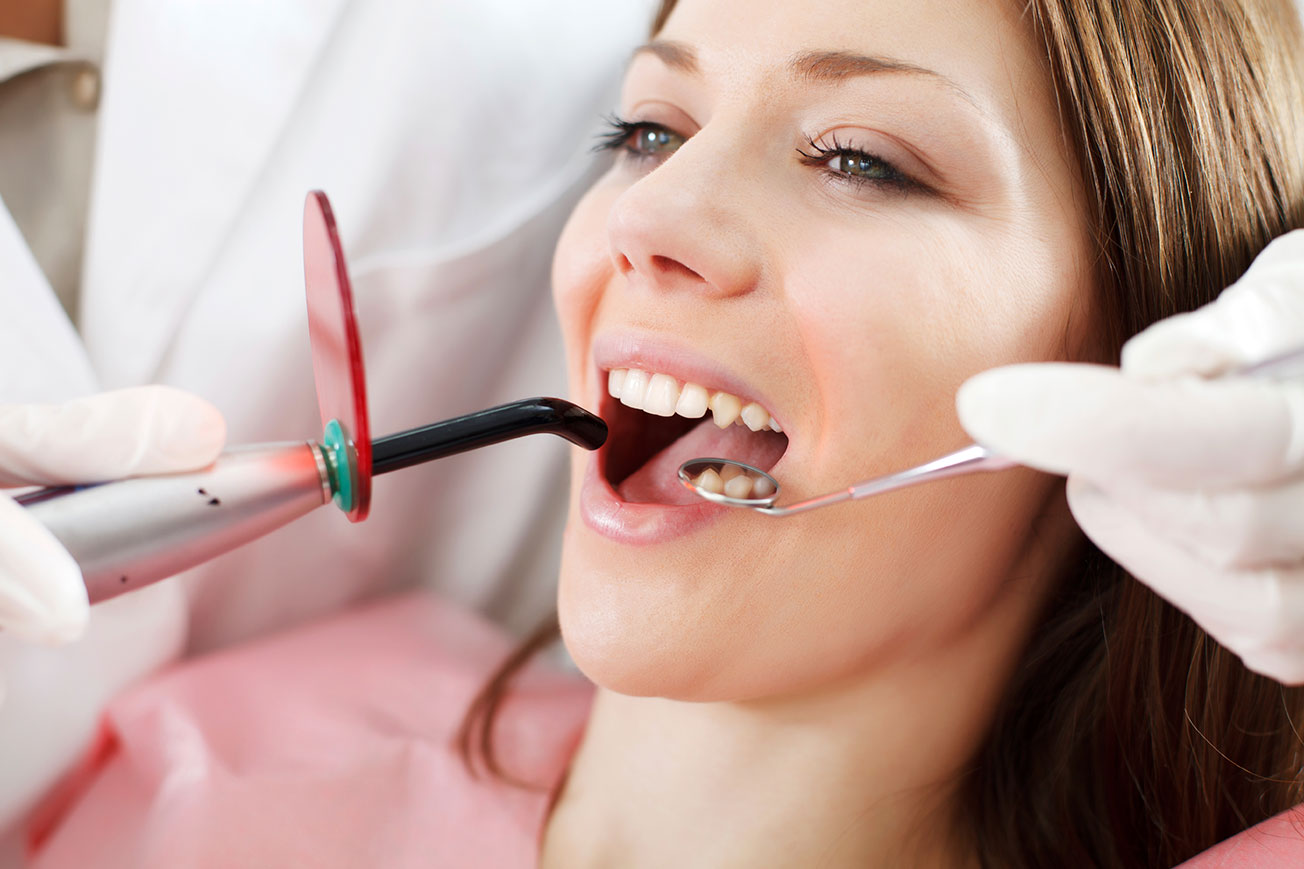
Research and Reviews:Dental medical fibers treatment
Keywords:medical fibers, treatment, Time:21-12-2015The word laser represents an elegant acronym as “Light Amplification by Stimulated Emission of Radiation”. It was demonstrated for the first time by Theodore Maiman in 1960 with various laser types (Nd:YAG, Er,Cr:YSGG, Er YAG, CO2)having corresponding wavelengths(1064nm, 2780nm, 2940nm, 10600nm) becoming available to dentists to address their needs for hard and soft tissue treatment procedures [1,2]. Interaction of lasers on soft tissue enables dry and bloodless surgery, minimal post operative swelling and pain where as lasers for hard tissue encourage efficient diagnosis of caries, cavity preparation and sterilization of root canal system. This article focuses on use of surgical lasers fibers in dental procedures.

History
The first medical fiber treatment introduced by Theodore Maiman was a synthetic Ruby laser [1,2]. Sognnaes, in 1964, reported glass like fusion and catering of enamel when subjected to 500-200J/cm2 of laser energy [3]. All the early researches were concentrated on ruby lasers. Nd;YAG laser were largely ignored during these years but ruby laser lost favor soon as it needed too much energy for effective dental hard tissue procedures and was reported to cause severe thermal damage to the pulp, collateral damage to adjacent hard and soft tissue due to scattered radiation. In 1964, carbon dioxide laser were introduced, wavelength suitable for clinical preparation of dental hard tissue were formed with excimer laser within ultraviolet range. Due to ablation and technical reasons, excimer laser showed a restricted ability in clinical use. Later in 1965, Goldman et al exposed a vital tooth for the first time to laser energy, which was painless procedure and only minor, superficial damage to the crown was observed [4]. In 1961 Weichman & Johnson tried in vitro to seal the apical foramen using high power infrared (CO2) laser [5]. In 1989, it was demonstrated that the Er: YAG laser can be used for preparing cavities in enamel and dentin without major adverse effects. Tokonabe et al,in 1999, said that cavity preparation with Er.YAG may result in ablation craters with chalky appearance on surface. In a study conducted to evaluate efficacy of Er;YAG laser revealed that lasers are equivalent to air rotar in preparing cavities.
Lasers in Operative Dentistry
In laser cavity preparation, intensive electromagnetic energy is used for ablation of tissues. Depending on wavelength of laser light, the ablative effects can be based on chemical or thermal effects. Erbium based system became widely accepted for preparation of dental hard tissues; with their specific ablation mechanism, they cause a microretentive pattern in walls of prepared cavity. This enhances the adhesion between composite material and the cavity. It also causes obliteration of dentinal tubules, which is also revealed by the studies in the last few years. However, removal of enamel and dentin by means of laser leads to thermal side effects. This fact was determined by Sterns (1964) who proved that until new ways of radiation production are introduced, laser will prevail with very strict limitations. The prognosis of Stern remained valid for more than a decade, until other lasers with a more favorable wavelength were introduced. The removal of enamel and dentin with no thermal side effects was possible once the superpulsed CO2 was introduced [6]. However during caries removal with this, cooling with water is indicated with no loss in laser intensity. In contrast to this Er:YAG, Cr:YSG lasers have advantage of reducing thermal effects [7]. Laser treatment is contact free and has advantages over rotary instruments such as, direct cooling of the area with water spray, absence of drilling sound, pressure, pain, temperature and anesthesia can be omitted.
Lasers in Endodontics
In endodontics, lasers have been used as adjuvant treatment in both - low-intensity laser therapy and high intensity laser treatment, to optimize the outcome of clinical procedures. Low-intensity laser therapy induces analgesic, anti-inflammatory and biomodulation effects at molecular level with photochemical responses improving tissue healing processes and less postoperative discomfort for patients. The clinical application of low-intensity laser in endodontic therapy has been considered useful in post pulpotomy (with the laser beam applied directly to the remaining pulp and on the mucosa toward the root canal pulp); post pulpectomy (with the irradiation of the apical region); periapical surgery (irradiating the mucosa of the area corresponding to the apical lesion and the sutures). For intracanal application, a fiber supported laser delivery system having appropriate diameter is required which is capable of delivering laser energy laterally [8,9]. In addition, disinfection will be achieved in contaminated root canals due to the bactericidal effect of thermal interaction [10,11]. High-intensity lasers such as Nd:YAG(neodymium: yttrium, aluminum, garnet), Er:YAG(erbium: yttrium, aluminum, garnet), Excimer, CO2(carbon dioxide) and diode have been recommended successfully as an adjuvant method in the endodontic treatment of contaminated canals to remove bacteria from the root dentinal surface as well as from deep dentinal layers, pulpotomies and pulpectomy.
Laser in Periodontics
Depending on absorption characteristics, mode of action and indications various lasers can be used in procedure such as gingivectomy, gingivoplasty, scaling and root planning. Usually Er; YAG, Nd;YAG , CO2 laser is used in removal of hyperplastic gingival tissues, as it is characterized by least post operative pain, absence of scarring etc [12,13,14] .Laser gingivectomy with Er: YAG laser is suitable for immunocompromised patients as it exhibits excellent anti bacterial effects. It is also used to remove concrement and plaque from root surface along with sufficient water cooling. While comparing lasers with conventional periodontal surgical procedures, reduction in plaque index, bleeding index, pocket depth and better reattachment was observed.
Laser in Orthodontics
The application of lasers in orthodontics depends upon potential advantage of lasers over conventional methods [15]. Classical methods of acid etching imply use of phosphoric acid (37%) as a gel or solution on the enamel surface. The time of action is of 15-60 seconds. The enamel appears matt, after washing and drying. The use of CO2, Nd;YAG, Er; YAG lasers is an alternative for enamel conditioning and has proved to be more effective than phosphoric acid. For laser etching the surface must be covered with accelerator and area will be irradiated until evaporation of accelerator. For esthetic reasons ceramic brackets are very frequently used in orthodontic treatment but when these brackets are detached from teeth it can cause enamel fractures, thus can be avoided by thermal detaching of brackets by CO2 and YAG lasers. Strobl and Tocchio presented the data about changes occurred during detaching of brackets with CO2, YAG lasers [16,17]. Studies also suggest that Er;YAG lasers fibers are widely used for enamel surface conditioning [18].
Lasers in Cosmetic Dentistry
Tooth discolouration is the change in colour of teeth as compared with adjacent teeth; it may be due to genetic malformations or genetic disorders. Bleaching of discoloured teeth has decreased need for invasive treatment. Bleaching is a chemical whitening teeth in which hydrogen peroxide, sodium perborate, chlorine etc are used. In office bleaching techniques may involve use of energy sources to increase rate of release of bleaching radicals. Different lasers produce different wavelengths, hence not all lasers are suitable for bleaching. Wavelength absorbed, scattered or transmitted through tooth structure cannot be used for bleaching as it will damage enamel dentin and pulp. KTP, Argon and Diode lasers are used in office bleaching.
It is also known as „soft laser therapy‟. It is based on the concept that certain low level of doses of specific coherent wavelengths can turn on or turn off certain cellular components or functions.
Related Articles
- An Innovative Device for Fractional CO2 Laser Resurfacing:Medical fiber Application
- Disposable bare fiber in oral and facial practice
- Topical Gel Application and Low Level Laser Therapy on Related Soft Tissue Traumatic Aphthous Ulcers
- WHY A CO2 DENTAL LASER?
- Interventional laser surgery for oral potentially malignant disorders: a longitudinal patient cohort study
- Er:YAG laser applications in dentistry
- Lasers for Dental
- Lasers Fibers for Pediatric Dental Patients
- The effects among three desensitive tooth methods
- Adjunct to Nonsurgical Treatment of Periodontal Disease
