
Adjunct to Nonsurgical Treatment of Periodontal Disease
Keywords:laser, treatment, medical, Time:25-11-2015The biofilms adhered on the internal and external walls of the periodontal pocket, the free biofilm and the possibility of a bacterial penetration through the epithelium to the underlying connective tissue can cause a gingival inflammatory reaction. This inflammation may progress with vasodilation, cellular migration and release of mediators, thus increasing the inflammatory response and perpetuating the disease. This situation makes microorganisms more resistant to drugs, which frequently are unable to reach the colonies protected by the matrix and by the presence of resistent bacteria (Donlan & Costerton, 2002). The inflammatory phenomena triggered by the bacteria and their waste products attracts macrophages that produce, among others, interleukin 1 (IL-1) and tumor necrosis factor alfa (TNF-┙), which have the ability to activate osteoclasts and produce bone resorption. TNF-┙ activates the adhesion molecules of the endothelial cells of the vessels, favouring the adhesion of monocytes and diapedesis. It also stimulates the arrival of T lymphocytes, which contribute with receptor activator of nuclear factor kappa B ligand (RANKL) to the bone, consequently favouring the bone loss (Kong et al., 1999). But this process is more complex as it needs some proteins such as nuclear factor kappa B (NF-kB), receptor activator of nuclear factor kappa B (RANK), RANKL and osteoprotegerin (OPG), among others, which may change the answer of the osteoclast precursors and therefore modify the osseous destruction.
Nowadays, antibiotics are used as complementary elements but its use should be restricted to the minimum due to the frequent development of resistances, and to the difficulty of maintaining stable and effective levels during a long period of time (Socransky & Haffajee, 2002). For this reason it becomes necessary the additional research of substances or techniques that can modify the pH, the oxygen concentration or the nutrient disposition of the dental plaque in order to modify the microflora of the biofilm. We also need to find systems able to interfere with the bacterial genetic signals and to modify the inflammatory response in the periodontal tissues.
2. Lasers employed in periodontics
Currently, different equipments of laser radiation (medical fibers,surgical fibers,medical fiber optics,laser fibers), are available in Periodontics, each one with particular features and diverse effects, making necessary the selection of the most suitable for each type of application (Table 1). Some of these lasers are effective in eliminating the residual calculus and detoxifying the radicular cementum (Er:YAG) (Aoki et al., 2004; Ishikawa et al., 2004; Schwarz et al., 2008); on the contrary others are unable to eliminate the calculus but can act over the soft tissues reducing the inflammation, as they modify the tissue oxidation systems and the cytokines which mediate in inflammation (Nd:YAG, diode) (Gómez et al., 2011). Although these effects over the tissues are difficult to evaluate clinically, they are guaranteed by molecular biology techniques. The results seem to be variable, but the investigation should help us to select the wavelength of the radiation, pulse duration, energy/power applied, pulse shape, repetition rate, time of exposure, sequence, type of wave, continuous (cw) or pulsed, type of applicator (cutout or rigid fiberglass), and other factors which can provide the desired objectives.
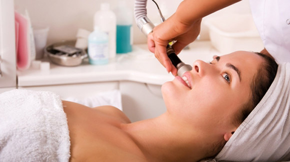
The therapeutic application of laser radiation can be clinically useful only if the appropriate instrumentation is available. Since the laser has been introduced into medicine, and so into dental discipline, a number of different applicators have been developed for day-to-day clinical use. The types of applicators most used in Periodontics are shown next. For instance, those with rigid fiberglass are set over handpieces. They should be used sliding them almost parallel to the radicular surface (Fig. 2), with a 20º angle in a coronoapical sense, as the perpendicular application produces damage in the cementum. Due to their size, the applicators with cutout fiberglass (Fig. 3) allow intrasulcular insertion and can reach deep areas. The displacement is also done in a coronoapical sense, outlining the whole radicular surface following the depth of the periodontal pockets, in the same manner as in a periodontal probing. Other rigid and thin applicators should be used with spiral or circular movements instead of coronoapically, in order to optimize results.
In Periodontics we need treatments to eliminate the plaque and calculus, and to eliminate and/or reduce the gingival inflammatory phenomena. We have to operate therefore over the soft and hard dental tissues. For this reason, the basic effects of periodontal lasers over these soft and hard tissues are presented next, in order to show the possibilities of this technology applied alone, and mainly in combination with SRP.
3. Lasers on dental soft tissues
The earliest clinical studies mentioning the application of lasers in the non-surgical treatment of periodontitis began in the early 1990s using a Nd:YAG laser with the development of flexible optical fibres. Since then, many studies have been carried out to evaluate the possible advantages of the use of lasers (Nd:YAG, diode (GaAlAs, InGaAsP), Er:YAG, Er,Cr:YSGG and CO2) with wavelengths ranging from 635 to 10,600 nm. Recently, systematic reviews have compiled different clinical and microbiological effects of different types of laser radiation used as monotherapy or adjunctive therapy compared with SRP. However, less information to demonstrate the anti-inflammatory effect of the laser radiation is available from the literature.
3.1 Nd:YAG laser radiation
Unlike other infrared lasers with a strong absorption by water, such as Er:YAG or CO 2, the wavelength of Nd:YAG laser presents a poor absorption by water, thus increasing scattering and infiltration of its energy into the biological tissues. The photothermal effects of Nd:YAG laser are useful in soft tissue surgeries. Thanks to its great penetration depth and thermogenesis properties, this type of laser produces a thick coagulation layer in the irradiated area, presenting a great haemostatic capacity, being efficient for the ablation of potentially haemorrhagic soft tissues (Perry et al., 1997, Romanos, 1994). There is little evidence to support the efficacy of Nd:YAG laser treatment as an adjunct to nonsurgical periodontal treatment in adults with periodontal inflammation. In the last decade, there are barely clinical studies published analyzing the clinical evolution and the inflammatory mediator levels in the gingival crevicular fluid (GCF) after irradiation with Nd:YAG laser in the affected sites in patients with chronic periodontitis. The results obtained in four clinical studies, performed by three different research groups, should be emphasized (Gómez et al., 2011; Miyazaki et al., 2003; Qadri et al., 2010, 2011). The overheating of the irradiated tissues and the consequent damage of the oral hard and soft tissues, could explain the limited support to this kind of laser radiation from the scientific community (Miserendino et al., 1994). For this reason, to avoid thermal damage, the irradiation parameters employed in these clinical studies were selected according to the results obtained in previous in vitro investigations, where potential morphological alterations of root surface irradiation were assessed under standardized conditions (Bader, 2000; Gómez et al., 2009). Concerning the evolution of the clinical parameters, the application of Nd:YAG laser both as monotherapy or as an adjuvant to scaling and root planing, did not offer significant advantages versus the treatment with ultrasonic devices, both at 8 (Gómez et al., 2011) (Fig. 4) and at 12 weeks (Miyazaki et al., 2003). Nevertheless, Qadri et al., in their split mouth trial, found better clinical results in the test side (SRP + Nd:YAG) than in the control side (SRP) during the long term follow up (up to 20 months) (Qadri et al., 2011). When analyzing the inflammatory mediators, Miyazaki, in a 12 weeks study, found a non statistically significant decrease of IL-1┚ in Nd:YAG group used as monotherapy in comparison with ultrasonic devices for the non surgical treatment of chronic periodontitis (Miyazaki et al., 2003). On the contrary, Gómez did find in a short term study (4 and 8 weeks) significant decreases both in IL-1┚ as in TNF-┙ when using Nd:YAG as an adjuvant to SRP versus SRP alone (Fig. 5). In this same study, the total antioxidant status (TAS) of the gingival fluid, gradually increased until the eighth week after the treatment with SRP + Nd:YAG, while it remained stable when the treatment consisted of SRP, being these differences statistically significant (Gómez et al., 2011). The total antioxidant capacity of the gingival fluid decreases in periodontitis as a consequence of the inflammatory lesion, and it recovers after non surgical therapy (Brock et al., 2004; Chapple et al., 2007; Tsai et al., 2005).
Related Articles
- Disposable bare fiber in oral and facial practice
- Lasers Fibers for Pediatric Dental Patients
- Clinical treatment applications of dental lasers fibers
- LOW LEVEL LASER THERAPY
- The TwinLight™ Concept in Dentistry Reviews
- Low level laser therapy in dentistry
- Er-YAG LASER AND DENTAL CARIES TREATMENT
- The ArF Excimer Laser on Human Enamel with Medical fiber
- Lasers for Dental
- The effects among three desensitive tooth methods