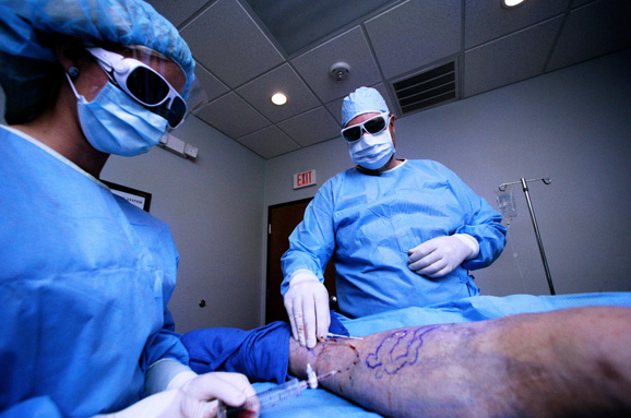
Endovenous Laser Ablation of the Small Saphenous Vein
Keywords:Endovenous, Laser, Time:19-07-2015Cohort study; Minimally invasive; surgical procedures
Abstract
Objective: To evaluate treatment of the small saphenous vein (SSV) by endovenous laser ablation.
Study design: A cohort study, occlusion of the vein and safety of the procedure was analysed prospectively.
Patients: 150 consecutive patients (169 limbs) were treated between August 2006 and January 2008 in an outpatient clinic setting. The average age was 57 years (range 23e87); 82% female; 31% had serious varicose disease (CEAP 3e6). Treated length averaged 23 cm (range 6e53 cm).
Methods: All patients underwent a standardised assessment comprising digital questionnaire, physical examination and duplex ultrasonography. The SSV was cannulated percutaneously under ultrasound control and perivascular local anaesthesia (tumescent) was injected. An 810 nm diode laser was used, delivering 70 J/cm. Three months post-treatment all patients received a duplex ultrasound of the treated vessel.
Results: Complete occlusion of the SSV after 3 months was achieved in 98% of the cases. Two patients (1.3%) had sural nerve paraesthesia. Six patients developed superficial thrombophlebitis. Serious complications did not occur.
Conclusions: Endovenous laser ablation for treating the incompetent small saphenous vein is a safe, effective and technically feasible technique.
Introduction
Endovenous laser ablation (EVLA) for varicose veins is a widely accepted form of treatment. Several studies have shown that endovenous ablation of the Great Saphenous Vein (GSV) achieves an outcome equivalent to surgical treatment by sapheno-femoral ligation and stripping.
The aim of our study was to assess whether EVLA of the small saphenous vein (SSV) can achieve the same results as in the GSV. The main advantages are that the procedure is minimally invasive, the entire vein is obliterated and accuracy of treatment is high, due to the continuous duplex visualisation of the vein during the procedure.
Surgical treatment of incompetence of the SSV usually involves SPJ ligation. Stripping of the SSV is not recommended in the Netherlands because of the risk of sural nerve injury, yet there is very little data to support this cautious approach.
Even in experienced hands sapheno-popliteal ligation is not always technically successful. This is mainly due to the diverse anatomic anomalies of sapheno-popliteal junction (SPJ) and its proximity to the tibial and sural nerves in the popliteal space (see Fig. 1). Rashid et al. have shown that ligation of the SPJ is not achieved in 30% of the cases, even if the junction is marked pre-operatively under ultrasound guidance. The incision made in the popliteal fossa is associated with wound healing problems and infection in 19-23% of cases. In theNetherlands, sapheno-popliteal ligationalone is recommended for the treatment of an incompetent SSV.6
Since conventional surgical treatment is associated with these adverse factors, we performed a prospective cohort study to analyse the safety and efficacy of EVLA in the SSV.
Materials and Methods
Study design
This cohort study included patients treated at the Centre for Phlebology Emmen, a specialised outpatient clinic for varicose veins, between August 2006 and January 2008. Following an interview using a standardised digital questionnaire and a physical examination, all patients underwent duplex ultrasound scan of the affected leg. The vascular surgeon, together with the dermatologist, decided on the best treatment modality for the patient. The severity of the venous insufficiency was graded following the C part of the CEAP (Clinical, Etiology, Anatomy and Pathophysiology) classification.
Patientswith incompetence of the SSVandclinical signs and symptoms were treated in preference by endovenous laser ablation. Those who preferred general anaesthesia or who had suffered reported severe phlebitis in their medical history (with evidence of intra-lumenal scars on ultrasound imaging)
were treated conventionally using surgical techniques.
Operative technique
The EVLA patients were treated in an outpatient setting. The whole procedure was performed under ultrasound guidance (Philips iU22 Ultrasound System, Philips Medical Systems, The Netherlands) by the vascular lab technician. Intra-lumenal access was achieved percutaneously using an 18 gauge needle at the most suitable distal point in the SSV. Factors determining this point were: the most distal point required to treat the incompetent segment of vein, the point just distal of the last tributary vein, or a change in diameter at which the distal part of the SSV was less than 3 mm in diameter.
A 5 Fr sheath was introduced over a standard J-wire. The laser fibre was inserted through the sheath and the tip was placed 2-3 cm from the SPJ. The main factors for determining this point were that the tip was placed beneath the fascia, and at a safe distance from the SPJ. Taking into consideration that the laser occludes at least 7 mm of the vein proximal to the tip of the laser fibre. An 810 nm diode laser (Delta 15 W, Diomed, MA, USA) was used with 14 W continuous mode, delivering 70 J/cm.
The whole procedure was performed under local tumescent anaesthesia (300 ml sodium chloride 0.9%, 20 ml xylocain 1%/adrenalin 1:200.000, 10 ml sodium bicarbonate 8.4%). No additional treatment for varices was undertaken contemporaneously, either by sclerotherapy or phlebectomy.
After treatment, all patients returned home with 35 mmHg compression stockings (Mediven Struva AG hip, Medi, The Netherlands) for 72 h. Patients were advised to walk regularly (at least 3 times daily 20 min) and prescribed diclofenac (50 mg three times a day) for 10 days postoperatively. Patients over 60 years of age or with symptoms received also omeprazole (40 mg once daily).
If the patient had further incompetent saphenous veins, these were treated by EVLA in the same session unless the patient preferred otherwise.
Follow-up
Six weeks after the procedure the patients were checked by an independent observer (physician assistant, who was not involved in the EVLA procedure) in the outpatient clinic for any remaining ecchymosis, pain, remaining symptoms and paraesthesia. This assessment allowed any further treatment by sclerotherapy or phlebectomy to be planned for a future date, if required. Three months after the procedure patients underwent duplex ultrasound scanning to assess the degree of occlusion of the vein and presence of thrombi in the deep venous system. Any occlusion less than the complete treatment length was scored as partially occluded.
All the data was collected prospectively in a Microsoft Excel database that was also used for statistical purposes.
Results
All procedures were performed by three vascular surgeons during an 18-month period. In total 169 limbs (150 patients) were treated. In the same time frame 11 patients were treated in the operating theatre under general anaesthesia by sapheno-popliteal ligated and phlebectomies. Two patients were not considered suitable for EVLA, because of intra-lumenal scarring after an earlier episode of thrombophlebitis, the 9 other patients preferred to be treated under general anaesthesia. At that time the operating theatre did not meet safety requirements to perform endovenous laser ablation, so they were all treated by the conventional surgical method.
The mean age of the patients treated was 57 years (23-87 years) and 82% were women. Twelve patients (7%) had had previous sapheno-popliteal ligation for the management of varicose veins. 31% had severe venous disease (CEAP 3e6). Mean SSV diameter was 6.6 mm (3.2-26.7 mm). Mean treated length was 23 cm (range 6-53 cm). The upper limit in the range was an incompetent Giacomini vein (see Table 1). 78 patients received concomitant treatment of the GSV in the same leg at the same time. 20 Patients received treatment of the GSV in the contralateral limb. One patient was treated for anterior accessory
saphenous vein reflux. 5 Patients had thrombophlebitis in the SSV (all were treated successfully).
Following the post-operative review at 6 weeks, sclerotherapy of saphenous tributaries was performed in 56 patients and one patient underwent phlebectomy. After 3 months duplex ultrasound scanning was performed in 150 limbs (89%). 19 Legs were lost to follow-up: 1 patient had died, 1 patient received underwent duplex ultrasound scanning, but the report was lost, and 15 patients (17 legs) did not attend for their appointment. This showed 148 completely occluded vessels (98%); 1 partially occluded (0.7%) and 1 not occluded (0.7%). No deep venous thrombosis was seen. Two patients (1.3%) complained about
numbness of the lateral lower leg and foot (the sural nerve); in one of these patients the paraesthesia was
completely resolved after two months. Six patients (6 limbs) had superficial thrombophlebitis which resolved spontaneously over time.
Discussion
Endovenous ablation by laser, radiofrequency or bipolar diathermy is the minimally invasive alternatives for obliterating the incompetent SSV.
Few case series on laser ablation of the SSV have been published (see Table 2). Proebstle was the first to write about treating the small saphenous vein with EVLA in 2003, 31 patients were treated, 2 patients (11%) had sural nerve paraesthesia and occlusion after 6 months of 100%.12 The second series was published by Ravi in 2006, 981 patients including 101 SSVs, sural nerve damage was not mentioned, of 37 SSVs the follow-up was 3 years with 84% occlusion.13 Theivacumar treated 68 SSVs with a follow-up of 6 months and reported 100% occlusion and transient numbness of the sural nerve in 4%.14 In 2007 Gibson describes a single patient group of 187 patients (210 SSVss) treated by EVLA, in which 96% was occluded after 4 months, 3 patients (1.6%) with numbness of the lateral malleolus.15
Park published the largest and most recent series (344 patients), 2% sural nerve paraesthesia was reported and an occlusion rate of 94% after 12 months in 108 patients.16

In the recent literature on endovenous laser ablation of the SSV the frequency of sural nerve paraesthesia was less than 2%.15,16
Very few publications in the recent literature have been published on the results and complication rate of the present surgical standard: ligation of the sapheno-popliteal junction.17 In one study assessing technical success of surgical of the junction, good or satisfactory results were achieved in only 59% of the cases, even if the insertion of the SSV in the popliteal vein was marked pre-operatively with ultrasound imaging.8 The frequency of sural nerve damage after surgical ligation of the SPJ and stripping of the SSV are highly variable, ranging from 0% to 21%.7
In conclusion, published data show that open surgical treatment of the SSV has a mediocre initial technical success, judged by duplex ultrasonography. EVLA of the SSV shows a high initial success rate. The main advantages are accuracy of treatment attributable to duplex guidance during the procedure and obliterating the entire vein instead of just dividing the sapheno-popliteal junction. Minimal post-operative morbidity was encountered since there was no requirement for an incision in the popliteal
fossa. Our series show that endovenous laser ablation of the SSV is a safe, effective and technically feasible technique.
At present surgery is the recommended treatment of reflux of the SSV in the Netherlands. Only clinical series of endovenous laser ablation of the small saphenous vein have been published thus far, with promising results. A randomized trial, comparing surgical treatment with endovenous laser ablation with at least two year follow-up, should be performed to assess the relative efficacy and complications of these treatments.
Related Articles
- Er-YAG LASER AND DENTAL CARIES TREATMENT
- Disposable bare fiber in oral and facial practice
- Laser Treatment for Failing Dental Implants
- Laser Fibers Application in Periodontics
- Medfibers:Diode Lasers for Periodontal Treatment
- Disposable bare fiber in oral and facial practice
- The clinical observation of semi-conductor laser treatment of hypersensitive dentine
- Mediastinal emphysema caused by a dental laser
- Global Industry Analysis on Medical Laser Technology Market, 2015 to 2021
- APPLICATION OF Nd–YAG LASER TREATMENT FOR ORAL LEUKOPLAKIA
