
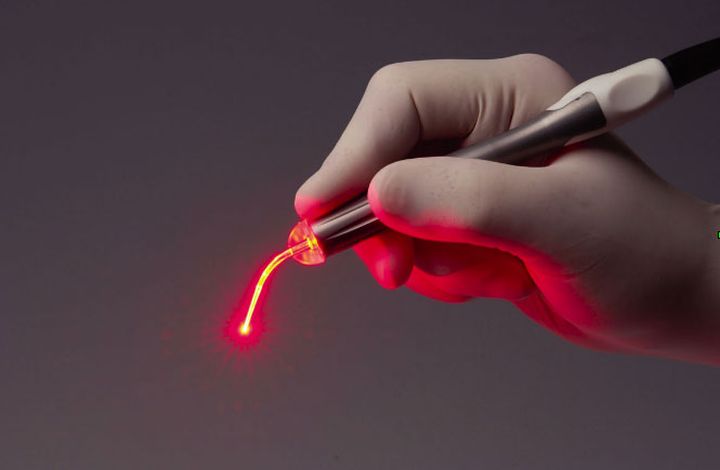
The ArF Excimer Laser on Human Enamel with Medical fiber
Keywords:medical fibers, surgical fibers, Time:16-12-2015The exposure of human teeth to a ruby laser caused the surface enamel to be more resistant to subsurface demineralization in vitro [7]. Further studies, however, revealed a severe irreversible damage to the pulp as a result of ex- cessive heating 14, 8-101. The use of other lasers such as the Nd:YAG laser (wavelength of 1,060 nm) and COz laser (wavelength of 10,600 nm) was therefore suggested [11,121. The effect of these physicochemical changes in tooth enamel confirmed increasing resistance to subsurface demineralization [7, 21-23, 27-311. There was an attempt to apply the CO, laser in a dental clinic replacing the drilling machine for caries removal, sterilization, dentin repair, and increasing resistance to caries [32,331. However, cracking and carbonization of the irradiated sur- face with damage of the pulp tissues were ob- served due to heat conduction during the laser tissue interaction [34-361. Laser application in dentistry was therefore limited to preventive den- tistry. Recent studies have focused on the Er:YAG, which is a pulsed Medical laser fibers with a wavelength of 2.94 pm. This laser was effectively applied to remove dentin and enamel with minimal damage of the surrounding tissue [37,38]. The rationale for us- ing the Er:YAG is principally concerned with the strong water absorption at this wavelength, which results in a micron-size absorption depth, and partially selective interaction with tissue having high water content. An alternate strategy for laser applications in dentistry is the use of excimer lasers. The ini- tial study with this laser was concentrated on the use of the XeCl excimer laser at 308 nm for the treatment of dental root canals [391. All the exci- mer lasers and especially the argon fluoride exci- mer laser at 193 nm are characterized by its nonthermal photoablation interaction with poly- mers [40,411, and, therefore, it seemed to be an effective solution for laser irradiation of tooth enamel without thermal damage to the pulp.
MATERIALS AND METHODS
Immedi- ately after extraction the teeth were carefully cleaned from soft tissue and blood under dissect- ing microscope using soft tissue paper to avoid any damage to the enamel surface. The tooth re- gions selected for laser irradiation were covered with sound enamel with no obvious caries or any other malformations. Approximately 10 parallel longitudinal (from mesial to distal) sections were made from each tooth, and the enamel surface was irradiated and analyzed as described below.
Irradiation of Enamel Surface With Excimer Laser
A Lamda Physik Model EMG 103 MSC ArF excimer laser was used with a wavelength of 193 nm and a pulse duration of 20 ns. The irradiation spot size was fixed to 1 mm in diameter by a metal mask. The laser beam was concentrated with a convex cylindrical lens and passed through this mask at average powers of - 25,30,35,40,45, and 50 mw with energy fluences ranging from 300 to 650 mJ/cm2 per pulse respectively. Laser beam power was measured with the DGX Laser Power/ Energy Meter, Ophir Ltd (Jerusalem, Israel). Seven sections were irradiated at each energy flu- ence. Each longitudinal section was irradiated in three regions: occlusal enamel, medial enamel, and cervical enamel, i.e., a total of 126 enamel regions were irradiated. The sections were fixed to a special holder that could be moved on an optic bench relative to the metal mask. They were ir- radiated at 10Hz for 4,000 pulses.
Etch Depth Measurement
A total of 252 etch depth measurements were made from 126 profiles of irradiated areas (two for each irradiated enamel region) using a scaled bin- ocular microscope
Examination of Irradiated Enamel With Scanning Electron Microscope
Human enamel specimens irradiated with the excimer laser at seven different energy flu- ences and an untreated specimen used as control were examined under JEOL 840 Scanning Elec- tron Microscope at 15KV. The samples were attached, with epoxy resin, to aluminium stubs then sputtered with - 200A thick layer of carbon.
RESULTS
The etch depth of each irradiated enamel area can be seen in Table 1. These data were an- alyzed using an analysis of covariance model. The results show that as the energy fluence increased so did the etch depth of the irradiated enamel (p = 0.0001) measured at three different regions: occlusal, medial and cervical (Fig. 1). No signifi- cant difference was observed in the etch depth between the three regions at the same energy flu- ences (p = 0.1457).
Scanning Electron Microscopy (SEM)
The surface morphology of occlusal enamel produced by a lOHz, 4,000 pulses laser treatment of intact human enamel at seven different energy fluences can be seen in Figure 1. The enamel was examined at the center as well as at the periphery of the irradiated areas. The control enamel surface, without laser ir- radiation, shown at low magnification (Fig. 1 con- trol(i)), is a relatively structureless layer of enamel with transverse wavelike grooves, periky- mata, believed to be the external manifestations of the striae of Retzuis. At higher magnification (Fig. 1 control(ii)) rod ends, cracks, and micro- pores were observed. The low 255 5 25 mJ/cm2 energy fluence laser treatment of enamel (Fig. la) produced very slight erosion observed under low magnification exhibited a delicate periodic surface roughening with a bright halo of - 100 pm width around the irradiated area.
Related Articles
- Lasers for Dental
- Dentine Hypersensitivity medical Laser fibers Therapy
- Study on the Influence of Semiconductor Laser Irradiated Time towards Dental Pulp and Dentin
- APPLICATION OF Nd–YAG LASER TREATMENT FOR ORAL LEUKOPLAKIA
- Effectiveness of diode laser as adjunctive therapy to scaling root planning in the treatment of chronic periodontitis
- APPLICATION OF Nd–YAG LASER TREATMENT FOR ORAL LEUKOPLAKIA
- LOW LEVEL LASER THERAPY
- Low level laser therapy in dentistry
- The TwinLight™ Concept in Dentistry Reviews
- Indications and limitations of Er:YAG laser applications in dentistry